Cam morphology
Instead of a smooth, round femoral head, there is a bony prominence that catches on the acetabular rim during hip movement. This is more common in men and in those who played high-impact sports during adolescence.
Pincer morphology
The acetabulum extends too far over the femoral head, creating an impingement as the hip joint tries to move in its socket. This is more common in women.
Mixed morphology
A combination of both cam and pincer features. This is the most common pattern in symptomatic patients.
Former high-impact athletes, dancers and highly flexible individuals
Those who played sports such as football, hockey, gymnastics, or martial arts during adolescence and those who partook in sports that had a high demand on hip flexibility.
Women with associated hip dysplasia
FAI and dysplasia frequently coexist.
Men with cam morphology
Often related to how the femoral head developed during adolescence.
Pain when sitting
FAI is characteristically worse with prolonged sitting (car journeys, desk work, cinema), because sitting places the hip in flexion (which is the position most provocative for hip impingement).
Deep groin pain
Felt inside the joint, not on the surface. Worse with hip flexion, rotation, and weight bearing activities.
Loss of internal rotation
The hip becomes progressively stiffer in internal rotation, noticeable during squatting, pivoting, or sitting cross-legged.
Sharp end-range pain
Deep squats, yoga, cycling with a low saddle position, and certain swimming strokes commonly trigger symptoms.
Clicking or catching
This occurs when a labral tear is present.
Clinical examination
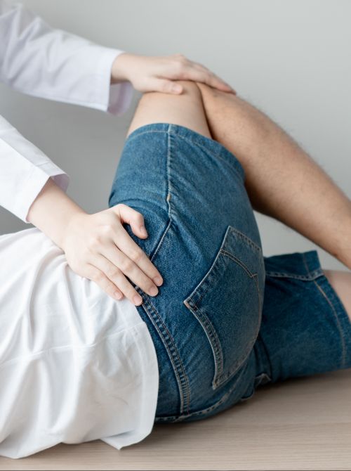
The FADIR test (Flexion, Adduction, Internal Rotation) reproduces symptoms by placing the hip in the position that causes impingement. This clinical test is positive in the majority of symptomatic FAI cases.
Hip X-rays
Specific X-Rays and measurements are taken to diagnose FAI. The X-Rays need to be properly positioned in order to get accurate measurements, not simply reported on a routine pelvis film.
MRI
An MRI scan is taken after injecting contrast into the hip joint. This is the gold standard for assessing labral tears and articular cartilage damage, and is significantly more sensitive than a standard MRI without contrast.
Physiotherapy
Involves strengthening the muscles around the hip, correcting movement patterns that may be contributing to the problem, and improving muscle control. This is a real treatment option in its own right, not simply a step to get through before surgery.
Injection treatment
Corticosteroid injection into the hip joint can provide meaningful symptom relief and confirm the hip joint as the pain source. Also useful to facilitate more effective physiotherapy engagement.
Hip Arthroscopy
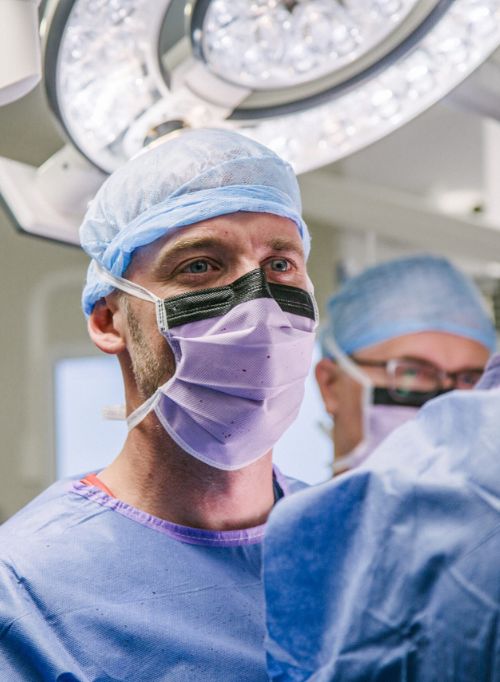
This is a surgical treatment for FAI in patients with preserved cartilage. During the operation, the abnormal bone can be reshaped (cam trimming, pincer correction) and the labrum repaired. It is performed entirely through keyhole incisions.
Hip replacement: the Kley Rapid Hip
When FAI has progressed to significant cartilage loss and osteoarthritis, a hip replacement is the appropriate treatment. Our consultants can assess your symptoms and scan results to identify the state of your cartilage and advise on the right treatment.
This is the single most important factor determining your treatment. Well-preserved cartilage points toward arthroscopy; advanced cartilage loss points toward replacement. Our consultants can organise the proper imaging and use the results to give you a specific, honest recommendation.
If you've been told you need a hip replacement but you're under 50 and haven't had an MRI arthrogram assessing your cartilage, it's worth seeking a specialist assessment to confirm that preservation surgery is genuinely no longer appropriate.