Delayed diagnosis is common
Research shows the average time to diagnosis is three to five years, with patients typically seeing four to eight clinicians in the process.
Symptoms overlap with other problems
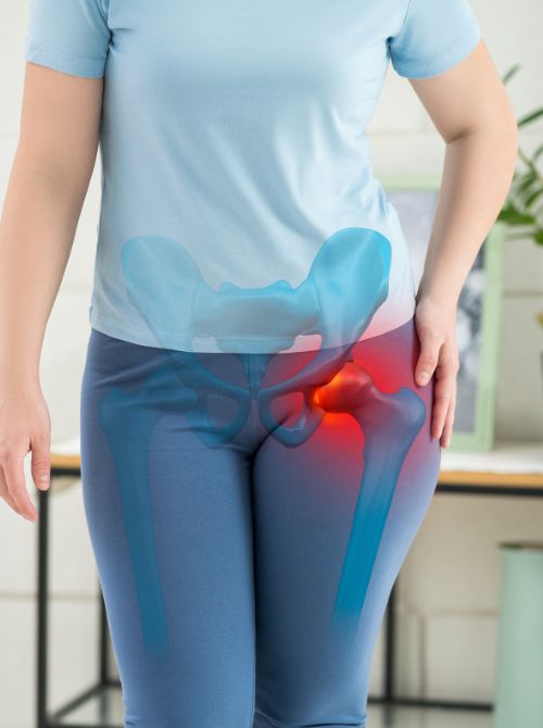
Groin pain, hip flexor tightness and instability are frequently mistaken for more common soft tissue diagnoses. Hip problems in younger women are often assumed to be soft tissue or gynaecological in origin.
Standard X-rays can miss it
Dysplasia often only shows up when specific angle measurements are taken, not on a routine X-ray report.
It's subspecialist territory
General orthopaedic and sports medicine practitioners aren't always specifically trained to recognise it, so it can be overlooked.
If you've been told "nothing significant on your scans" after months or years of hip symptoms, a specialist hip assessment with proper imaging may reveal a diagnosis like this that has been missed.
Prevalence
Affects approximately 3–5% of the population to some degree.
Gender
Considerably more common in women.
Birth factors
Disproportionately affects first-born children and those born breech (feet first).
Genetics
Has a recognised genetic component.
Deep groin pain
An ache inside the joint that worsens with activity and improves with rest.
Instability or giving way
A sensation of the hip slipping, catching, or feeling unreliable.
Pain with specific activities
Climbing stairs, getting in and out of cars, pivoting, running, yoga, or dancing.
Clicking or catching
Often related to a labral tear, a common consequence of dysplasia.
Night pain
As the condition progresses, pain at rest becomes more common.
X-ray with angle measurement
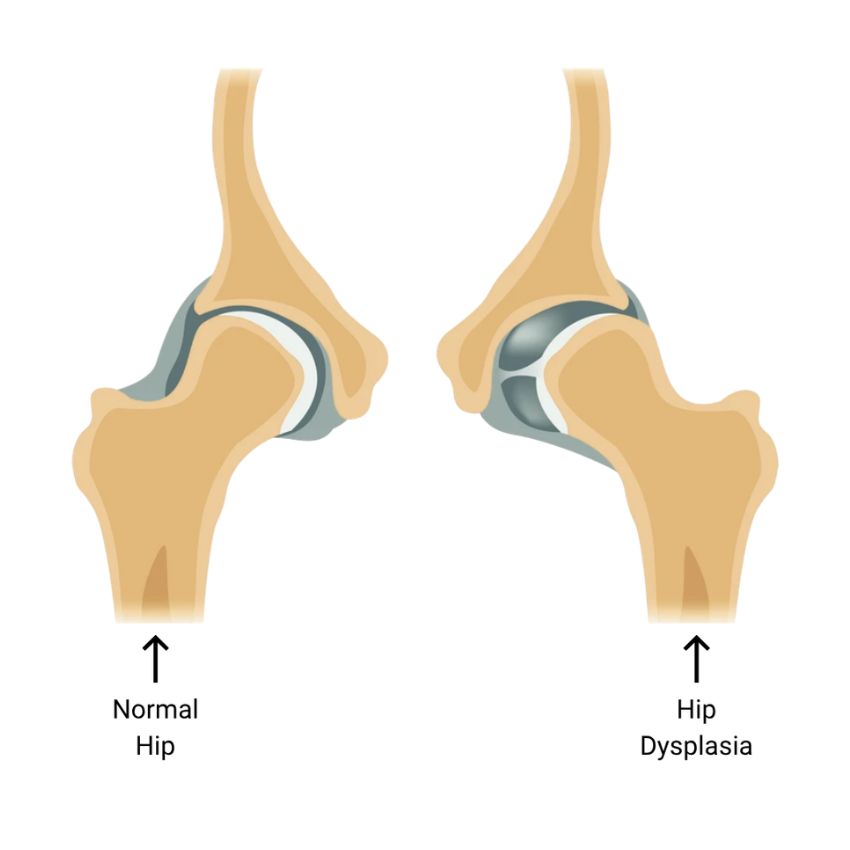
Measures the angle of the hip socket. A reading below 20 degrees (the lateral centre-edge angle, or LCEA) indicates dysplasia. A second X-ray, from a different angle, checks coverage at the front of the socket.
MRI arthrogram
Assesses the labrum for tears and the cartilage for early damage. It's far more sensitive than a standard MRI for picking up these issues.
CT scan
Provides a detailed 3D view of the socket's shape and angle, used when planning surgery.
Physiotherapy
Targeted strengthening of the hip stabilising muscles.
Activity modification
Adjusting movement and load to reduce symptom flare-ups.
Injection treatment
Corticosteroid or hyaluronic acid injections can reduce symptoms and slow progression in milder cases, or act as a bridge while surgical options are planned.
Hip preservation surgery: Periacetabular Osteotomy (PAO)
The definitive surgical treatment for significant dysplasia with preserved cartilage. Controlled cuts are made around the acetabulum, allowing the socket to be repositioned to provide better femoral head coverage, then fixed in place. OI provides complete assessment for PAO and works with specialist PAO surgeons where this is the recommended treatment.
Hip replacement: the Kley Rapid Hip
When cartilage damage has progressed to significant osteoarthritis, a hip replacement becomes the appropriate treatment. Hip replacement in dysplastic anatomy is more technically demanding than a standard replacement, as the abnormal socket anatomy must be managed carefully to achieve correct component positioning. The Kley Rapid Hip's pre-operative computer simulation specifically addresses this, planning component positioning in advance of surgery for every case.
The right treatment depends on how much articular cartilage remains. Our consultation, with specific hip imaging, will give you a clear, honest answer about which treatment is appropriate for your stage of disease.
Left untreated, hip dysplasia will progress. Studies suggest it's an underlying cause in a significant proportion of hip osteoarthritis cases diagnosed in women before the age of 55. Joint-preserving surgery (PAO) only remains an option while enough cartilage is left. Once enough cartilage has been lost, a hip replacement becomes the only option. A patient with preserved cartilage at 34 has choices that a patient with bone-on-bone contact at 44 doesn't. Catching this early can genuinely change what treatment is possible.